|
|
 |
GLUCOSE METABOLISM
 Glucose
metabolism
Glucose is virtually the only energy substrate which the
brain can use. Free fatty acids, used by most other
tissues when glucose is in short supply, are excluded
from the brain by the blood-brain barrier. The brain extracts 6.6 mL of O2
from each 100 mL of cerebral blood and returns 6.7 mL of
CO2.
Thus the respiratory quotient (RQ) of the brain is
approximately 1.0, indicating carbohydrate utilization
only. Brain glucose consumption is normally about 10 mg
per 100 ml., accounting for almost 75 percent of the
liver's production and further attesting to the brain's
heavy dependence on glucose.
Adenosine triphosphate (ATP), produced by the
metabolic degradation and oxidative phosphorylation
of glucose, is the useful energy currency in brain
tissue. About 85 percent of the circulating glucose
extracted from the cerebral arterial blood is
converted to CO2 via the tricarboxylic
acid (TCA) cycle, while 15 percent is converted to
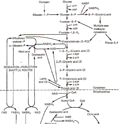
lactic acid. The general scheme for glucose
metabolism in the brain is similar to that in other
tissues and is illustrated in Fig.1. The
enormous ATP requirements of the brain are partly
due to neurotransmitter synthesis, release, and
reuptake as well as intracellular transport and
complex synthetic mechanisms. But undoubtedly the
greatest percentage of ATP is utilized to power the
ion pumps which restore membrane potentials,
enabling neurons to maintain their excitability.

Fig-1
 EFFECTS OF GLUCOSE DEPRIVATION
In
the healthy normal functioning brain, glucose is the only
substrate utilized for energy metabolism. Thus hypoglycemia
presents the brain with a very serious problem. While most other
tissues can shift to utilizing free fatty acids (FFA) as an
alternative energy source when glucose is lacking, the brain
cannot because they are excluded by the blood-brain barrier.
While there is some evidence that the brain can utilize
β-hydroxybutyric acid for
energy metabolism when glucose levels are low or when fats are
being mobilized for energy metabolism throughout the rest of the
body, the brain could never supply its high energy demands by
this method alone in the absence of glucose. Thus the brain is
dependent on an uninterrupted supply of blood-borne glucose to
energize its cells.
Decreases in blood glucose bring on disturbances in cerebral
function. Depending on the level of hypoglycemia, these changes
range from mild sensory disturbances to coma. At blood glucose
levels of 19 mg per 100 mL or
below (normal is 60
to 120 mg per 100 mL), a mentally confused state occurs. Brain O2
utilization falls to 2.6 mL per 100 g per minute (normal, 3.5 mL
per 100 g per minute) and glucose utilization drops as well.
Coma commences when glucose levels fall to 8 mg per 100 mL.
Epinephrine can be effective in reversing the effects of
hypoglycemia by promoting liver glycogenolysis. However,
attempts to solve the problem by substituting other carbohydrate
metabolic substrates have been largely unsuccessful, with the
single exception of mannose. This is the only monosaccharide
other than glucose which the brain appears to utilize directly.
It crosses the blood-brain barrier and directly replaces glucose
in the glycolytic pathway. However, its normal level in the
blood is too low to be of any real help in reversing the
cerebral effects of hypoglycemia. Unless reversed quickly,
comatose levels of prolonged hypoglycemia will bring on necrosis
of cerebrocortical cells and (to a lesser extent) other brain
regions as well.
|
 |
 |
|

Prof. Munir Elias
Our brain is a mystery and to understand it, you
need to be a neurosurgeon, neuroanatomist and neurophysiologist.

neurosurgery.tv

Please visit this site, where daily neurosurgical activities are going
on.

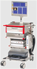
Inomed ISIS IOM System
|
|
|
|