|
|
 |
DESCENDING MOTOR PATHWAYS
Sherrington
called the motor neuron the final common pathway. All the subtle
signals converging from several descending tracts as well as
afferent input from the periphery are somehow integrated on the
motor neuron, which subsequently conducts the appropriate signal
out to the muscle. Because so many different pathways converge
on the motor neuron, the contribution of any single tract to the
final motor act is extremely difficult to determine.
Several
descending pathways have been shown to effect changes in the
activity of motor neurons. The anatomical courses of these
pathways have been extensively studied from their origins in
various areas of the brain to their synaptic contacts with the
motor neurons. The precise physiological roles of these pathways
have been studied but the information is limited because of
several factors. Principal among them is the fact that most of
the work has concerned the motor neurons innervating hind limbs
of the cat. Studies on primates have been continuing, but a big
problem is the somewhat suspect attempt to wed the
neurophysiology of the cat's movement performance to the
neuroanatomy of the human's.
Another
problem lies in the fact that a common tool for studying the
function of nerve pathways is electrical stimulation. While
there seems to be little alternative to this procedure, the
meaningfulness of artificially induced volleys of impulses is
questionable when one considers that tile natural influences on
motor neurons are spatially and temporally varied and probably
achieve their effects by virtue of a pattern of impulses rather
than a repetitive volley. Recent attempts have been made to
study the neurophysiology of movement by recording neuromuscular
potentials accompanying spontaneous movement. This is certainly
a desirable approach but is also limited by the fact that even
simple body movements are neurally very complex. Thus attempts
to relate the anatomical and physiological events associated
with these movements are difficult and hard to interpret.
Nevertheless, much has been learned concerning the role of the
nervous system in such activities as walking, running, and the
regulation of postural movements. It now appears that there are
"pattern generators" or "prewired" groups of neurons within the
central nervous system producing a wide variety of basic motor
programs. "Command" neurons activate these pattern generators
when a particular movement is called for. Here, we will examine
some of these pattern generators as well as the role of the
brain and its descending pathways in initiating and regulating
movement.
 UPPER AND LOWER MOTOR NEURONS
Electrophysiological studies have shown that the motor cortex
resembles a map showing a distorted image of the body turned
upside down and reversed left to right. Some motor pathways to the skeletal
musculature of the body arise directly from cells within the
cerebral motor cortex, while others arise from subcortical
areas of the brain and brainstem.
Neurons
that originate in the cerebral motor cortex. the cerebellum, or
various brainstem nuclei that send axon, into the brain stem and
spinal cord to activate cranial or spinal motor neurons are
called upper
motor neurons, Those cranial and spinal motor neurons which
actually innervate muscles are the lower motor neurons.
The latter include the
alpha and gamma motor neurons of spinal nerves. Upper motor
neurons are found entirely within the CNS, while the fibers of
lower motor neurons are part of the PNS.
Upper motor
neurons are clustered together to form descending tracts in the
brain and spinal cord. Such tracts are commonly named according
to their site of origin and the region of their distribution. An
example is the corticospinal tract, which originates in the
cerebral cortex and is distributed to the spinal cord. Another
is the rubrospinal tract. which originates in the red nucleus
(nucleus ruber) of the midbrain and is distributed to the spinal
cord. The lower motor neurons of spinal nerves are
somatotopically organized in the anterior horn of the spinal
cord gray matter. In general, those innervating the distal limb
musculature are located in the lateral aspects of the anterior
horn, while those innervating proximal limb muscles are found in
the intermediate region, The most medial group of motor neurons
innervates the musculature of the appendicular and pelvic
girdles.
PATTERN
GENERATORS AND THE CENTRAL PROGRAM FOR MOVEMENT
Upper motor
neurons don't simply stimulate lower motor neurons and produce
movement. The highly skilled and coordinated movements of which
humans are capable would seem to require a more complex and
involved system. While little is known of the highly involved
and integrated activity which occurs in the brain's neural
circuits during even a simple body movement, it now appears that
highly coordinated and very complex systems of interneurons
regulate the precise timing and sequencing of muscle activity
which is observed in such movements. There is also increasing
evidence that groups of interneurons cause specific patterns of
impulses to fire in the lower motor neurons associated with a
given coordinated movement. The central theory is that these
interneurons form pattern generators within the CNS which
produce the basic motor program. At the spinal cord level the
pattern generator is composed of a set of local control centers
located in the gray matter. There are neurons within these
centers which coordinate muscular synergies and generate timing
signals. Command neurons activate these pattern generators when
a particular coordinated movement is required. The result of
such activation is that the lower motor neurons fire in a
properly sequenced and timed pattern to produce a coordinated
movement.
Identification of specific command neurons for a particular
human movement is a difficult process and a speculative one at
best. It may be that upper motor neurons from the brain and
brainstem function in this respect for voluntary movements and
reflex postural adjustments. In some invertebrates, however,
the activation of a single interneuron is sufficient to excite
an entire coordinated muscle behavior. For example, stimulation
of the giant axon of the crayfish produces a coordinated tail
flip which propels it away from the stimulus. Similarly,
stimulation of the Maunther cells of teleost fishes produces a
tail flip propelling the fish away from the stimulus. These
cells are part of the reticulospinal tract neurons in the fish
and apparently serve as command neurons that activate the
pattern generator which carries the motor program for tail flip.
It is
presumptuous to assume that all movements proceed in accordance
with pattern generators and prewired motor programs.
Nevertheless, it may be that certain basic coordinated movements
of the limbs and trunk may proceed in a very general way under
the influence of such programs, while the initiation and fine
tuning of the movement requires input from descending and
sensory pathways. The upper motor neurons of some descending
motor pathways no doubt serve as command neurons for certain
movement patterns. Variations in the discharge patterns of these
neurons determines the variability of the programmed response.
There is evidence that a change in the firing frequency of
certain command neurons leads to a change in the intensity of
the response. If the coordinated movement involves a postural
system, altering the firing rate alters the magnitude of
postural adjustment. If a locomotor system is involved, the
frequency of the movement cycle will vary with changes in the
frequency of command
neuron firing. Other command neurons produce the same motor
pattern regardless of their firing rates. Their role seems to
be simply turning the program on and off. Still others may
regulate the magnitude of the programmed response. Most of the
vertebrate work involving motor programs has dealt with locomotor activity in the cat. Perhaps we can get a feel for the
intricate features of such programs by an examination of this
work.
Locomotion
in the Mesencephalic Cat
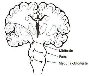
Removal of
the telencephalon (cerebral hemispheres) and the rostral portion
of the thalamus in an acute cat preparation remarkably leaves
the animal with practically normal locomotion. It can walk
spontaneously on its own and it can be forced to run by
electrical stimulation of a region in the subthalamus called the
subthalamic locomotor region (SLR). However, spontaneous walking
movements cease in acute preparations where the brainstem is
sectioned caudal to the subthalamus but just rostral to the
midbrain. This is a mesencephalic preparation, meaning the
highest intact brain component is the midbrain or mesencephalon.
There are several advantages to the study of locomotion in such
a preparation. Perhaps most important is that relatively normal locomotor movements can be initiated by the electrical
stimulation of an area in the tectum of the midbrain called the
mesencephalic locomotor region (MLR). Such a cat preparation can
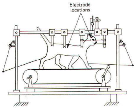
also be placed on a treadmill to facilitate natural movements
while its head is fixed in a stereotaxic apparatus enabling the
experimenter to conveniently stimulate various brain stem areas
and observe the
results (Fig-1).
 |
 |
| Fig-1 |
Fig-2 |
Walking and
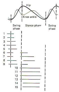
running movements in a mesencephalic cat are similar to those
observed normally. A single step cycle is accomplished when a
limb touches down, lifts off and moves forward, and then touches
down again. The step cycle is composed of a stance phase (limb
in contact with the ground) and a swing phase
(limb lifted off and moving forward). Muscle group activity
during the step cycle proceeds in a logical order. At the end of
the stance phase, when the limb is at its most caudal position,
the flexors become active, lifting the leg and initiating the
swing phase. During the swing phase, the relaxation of the
flexors combined with onset of extensor activity and inertia all
propel the limb forward. Because the extensors begin to contract
prior to the stance phase, the limb is able to support the
weight of the body as the limb touches down. Extensor activity
continues throughout the stance phase until just at the end,
when it begins to diminish and is replaced by increasing flexor
activity preceding another step cycle (Fig-2).
Increasing
the stimulation of the MLR causes an increase in the stepping
frequency of the mesencephalic cat. However, this increased
frequency is apparently due to an increased muscular force
moving the treadmill faster with the rate of stepping increasing
indirectly to keep up with it. The increased force is apparently
due to the increased recruitment of more alpha motor neurons and
motor units rather than to any increase in the firing rates of
the currently active units. Thus MLR stimulation directly
increases the level of muscular force and indirectly the
stepping frequency. If the level of MLR stimulation is held
constant and the treadmill is either speeded up or slowed down
by the experimenter, the stepping frequency of the
mesencephalic cat will speed up or slow down accordingly.
It is very
interesting to note that while stepping is a complex process
involving a repetitive sequence of muscular contractions and
relaxations with very precise timing, all that is necessary to
get it started is to stimulate the MLR in the mesencephalic cat
or the SLR in the subthalamic cat. Thus it seems likely that
stepping is an automatic process with a central program
controlled by a pattern generator in the CNS. Stimulation of
the MLR and SLR activates this program and can in fact vary its
intensity. The pattern generator for stepping with the hind
limbs of the cat appears to reside in the spinal cord. In
chronic cat preparations where the lower thoracic spinal cord
was completely sectioned shortly after birth. the animals are
capable of a full variety of stepping gaits in accordance with
the speed of the treadmill. Thus it seems likely that the
pattern generator resides in the spinal cord, at least for hind
limb movements. Transection of the spinal cord at a high enough
level to include the forelimbs (high cervical) does not
ordinarily allow for satisfactory locomotor movements. and thus
has not been adequately evaluated in this regard.
Input to
the Pattern Generator
Signals
arrive at the pattern generator in the spinal cord both from the
periphery and from supraspinal levels. The Ia afferents from
muscle spindles monosynaptically stimulate homonymous alpha
motor neurons and thus influence the activity of an ongoing
motor program. Similarly, signals arriving at spinal cord
interneurons from supraspinal levels via upper motor neurons
also exert an influence over the performance of a motor program.
It is
important to note that the stretch reflex is not always
productively useful at all times during the step cycle. Thus it
is not surprising to find that the sensitivity of the reflex is
varied cyclically with the step. It is "tuned in" when the
reflex is useful and "tuned out" when activation of the reflex
would be counterproductive to a particular phase of the step
cycle. Muscle spindle sensitivity can be controlled by the
pattern generator since it can apparently direct the timing of
both alpha and gamma motor neuron firing. Thus during the phase
of the stepping cycle when a muscle is passively stretched (i.e., the gastrocnemius at the end of the stance phase) the
sensitivity of its muscle spindles is decreased. This prevents
the stretch reflex from activating muscle contraction during the
"wrong" phase. Thus the spindles are "tuned out" when the muscle
is passively stretched and "tuned in" again when the muscle
becomes active during the step cycle. Let's now examine the
descending motor pathways which
influence motor activity.
DESCENDING
MOTOR PATHWAYS
|
Descending
motor pathways are defined as those which initiate or modify
performance and which originate in the brain. While several
tracts have been anatomically identified and physiologically
studied, it is still speculative to assume that we fully
understand what contribution any given tract makes to a
spontaneous movement. To electrically stimulate a descending
tract, observe a movement response, and then assume that the
observed response represents the function of the tract is surely
dangerous. The tract may have other, perhaps more important,
functions to perform which are not observed in the movement. Or
possibly the participation of the tract in a spontaneous
movement of the same kind may be of considerably different
magnitude. Nevertheless, stimulation of descending motor
pathways does produce activity in groups of flexor and extensor
muscles. Examination of these effects may give us valuable clues
as to the role of these pathways in normal spontaneous movement.
The
Corticospinal Tracts
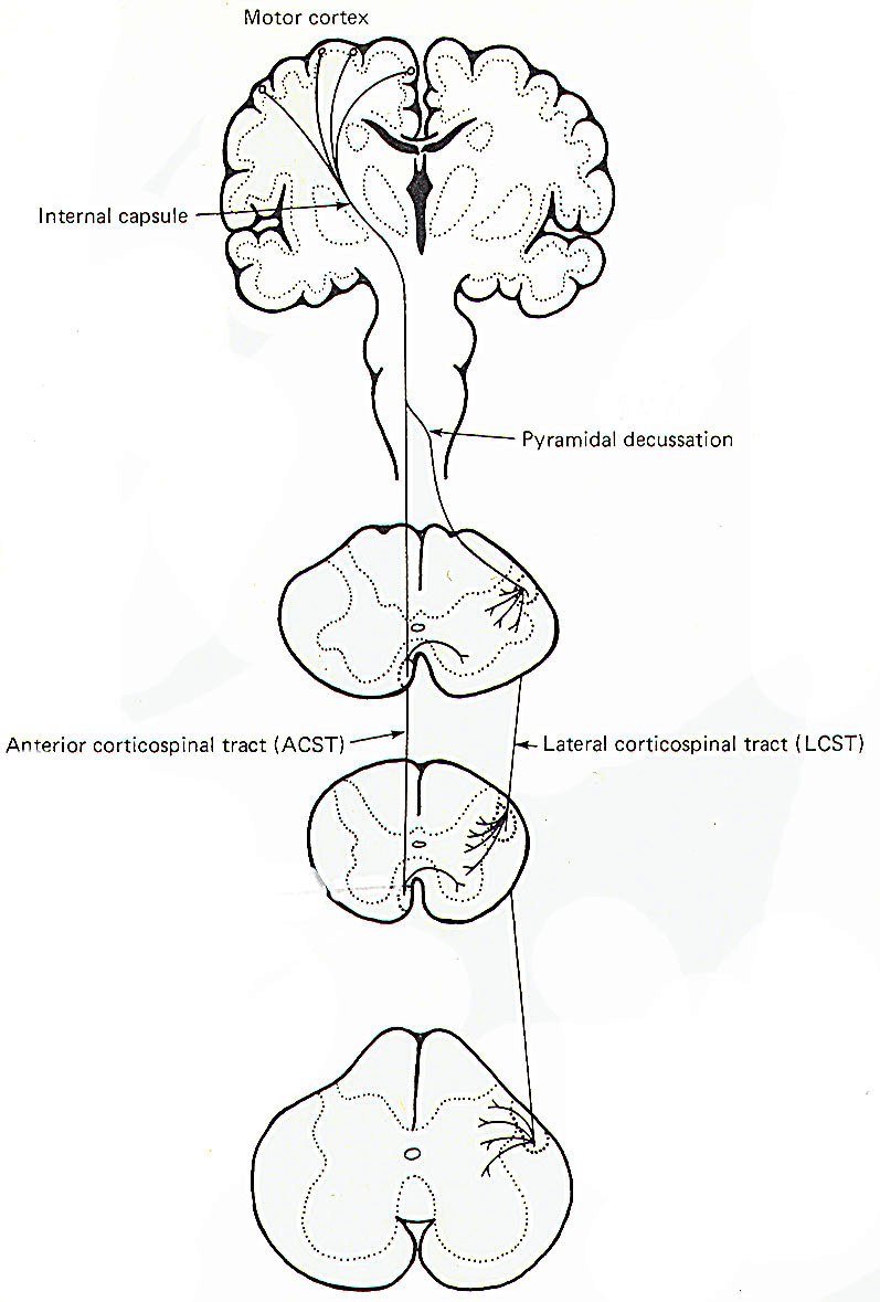
The
corticospinal tracts are often called the pyramidal tracts
because they form pyramid-shaped enlargements on the anterior
surface of the medulla. They are primarily concerned with
controlling skilled movements of the distal extremities and, in
particular, facilitation of those alpha and gamma motor neurons
which innervate the distal flexor muscles (Fig-3). There is
also evidence that they inhibit distal extensor muscles. The upper
motor neurons of these tracts originate in the precentral gyrus
of the cerebral cortex. From here their fibers pass without
synapsing all the way to their terminal destinations in the
spinal cord gray matter. After leaving the cortex, the fibers
descend through the posterior limb of the internal capsule, through the middle portion of the cerebral
peduncles to the basilar portion of the pons, and on into the
medulla oblongata where they form the medullary pyramids. Most
of the fibers (85 percent) cross over (decussate) to the
opposite side in the pyramidal decussation, where they continue
to descend in the lateral funiculus of the spinal cord as the
lateral corticospinal tract (LCST). The tract descends all the
way to sacral levels with fibers continually leaving it in order
to synapse on interneurons in laminae IV, V, VI, VII, and VIII.
Some even synapse directly on alpha and gamma motor neurons in
lamina IX (Fig-3). Those corticospinal fibers which do not
decussate in the medulla continue descending on the same
(ipsilateral) side of the cord and become the anterior
corticospinal tract (ACST). This tract does not extend below the
midthoracic level. Fibers leave the tract at various levels to
cross over in the anterior white commissure to synapse on
interneurons in lamina VIII.
Corticospinal Stimulation of Motor Neurons
Electrical
stimulation of the cortical areas from which the corticospinal
tracts arise excites many more motor neurons to distal forelimb
muscles in the baboon than it does motor neurons to proximal
muscles. In fact, proximal limb muscles are frequently not
activated at all by cortical stimulation. The more dextrous the
distal muscles are, the greater effect the corticospinal tracts
seem to have on their activity. Following cortical stimulation,
larger EPSPs are seen in the motor neurons to skilled distal
flexors than are observed in proximal muscle motor neurons.
Destruction
of the Corticospinal Tracts
Studies have shown that following
complete bilateral pyramidal tract section in monkeys, they are
still able to perform a wide range of activities using the body
and limbs and are able to walk and climb in a normal manner.
Their principal and most dramatic shortcoming is in their
ability to perform skillful manipulative tasks with the fingers
and hands. In similar tests of manipulative skills in monkeys
with unilateral pyramidal
tract sections, it was found that skilled movements in the
affected hand were dramatically reduced relative to the normal
hand. However, the animals were still able to move the whole
limb around the joints of the pectoral and pelvic girdles with
no trouble and they showed no difficulty in performing combined
movements of the limbs and the body. Thus it seems probable that
the corticospinal system is directed effectively to facilitating
movements requiring skill and dexterity of the distal
musculature. |
 |
| Fig-3 |
|
The
Corticobulbar Tract
This tract
is composed of fibers originating in the precentral gyrus of the
lower quarter of the motor cortex. The descending fibers leave
the motor cortex and pass through the posterior limb of the
internal capsule just anterior and medial to the corticospinal
tract fibers. From here they continue on through the cerebral
peduncles just medial to the corticospinal tract fibers to
terminate in the motor nuclei of cranial nerves III and IV in
the midbrain; V, VI. and VII in the pons; and IX, X, XI, and
XII in the medulla. The corticobulbar fibers from one side of
the brain project to the motor nuclei on both sides of the
brainstem (Fig-4).
|
 |
| |
Fig-4 |
|
The
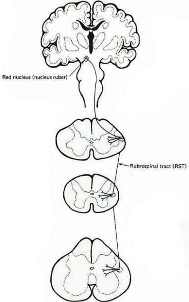
Rubrospinal Tract
The fibers
of this tract originate in the red nucleus (nucleus ruber) of the
midbrain. They cross over near their point of origin and descend
contralaterally in the lateral funiculus of the cord adjacent to
the lateral corticospinal tract (Fig-5). Before leaving the brainstem, some fibers of the tract enter the reticular
formation. As the tract descends through the spinal cord, fibers
leave it and synapse on interneurons in laminae V. VI. and VII.
Cells in the posterior portion of the red nucleus give rise to
axons influencing motor neurons of the neck and upper limbs.
while fibers from the anterior portion descend to lumbar levels
where they influence lower limb muscles.
Ablation
studies in which the tracts are experimentally cut have shown
that the corticospinal and rubrospinal tracts have somewhat
similar effects on the motor neurons. When the rubrospinal
tracts of monkeys were damaged on top of earlier pyramidal tract
sections. the loss of skilled control of the distal musculature
became even more severe and yet there was little or no loss of
control in the proximal muscles. Lawrence and Kuypers concluded
that a laterally placed group of descending fibers, which they
called the lateral system (corticospinal, rubrospinal, and
possibly other tracts), is primarily concerned with delivering
cortical control to the distal limb musculature. Independent
electrical stimulation of the intact rubrospinal tract
facilitates flexor and inhibits extensor alpha and gamma motor
neurons to the distal muscles.
Considering
that the red nucleus receives input from the same area of the cerebral
cortex as the corticospinal tracts, the similarity of their
actions may not be too surprising. The red nucleus also receives
input from the deep cerebellar nuclei and possibly the basal
nuclei as well. Nevertheless, as previously pointed out, the
reader should bear in mind that ablation and electrical
stimulation studies give us an incomplete picture of the
function of a descending, or for that matter any, tract in the
central nervous system. Further, whatever information is
obtained relates to the unnatural experimental situation and
not necessarily to normal function in the intact spontaneous
animal.
|
 |
| Fig-5 |
|
 The
Reticulospinal Tracts
The
Reticulospinal Tracts
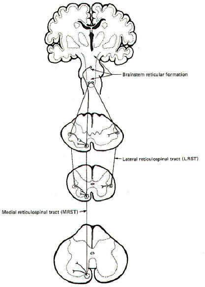
The
reticular formation is an indistinct group of cell bodies
clustered in the core of the brainstem. They don't form distinct
nuclear groups like those found elsewhere in the CNS. The
reticulospinal tracts represent groups of fibers which originate
in the reticular formation and descend into the spinal cord
(Fig-6). Those fibers which originate in the medullary
reticular formation show both a crossed and an uncrossed
component which descend in the lateral funiculus of the spinal
cord as the lateral reticulospinal tract (LRST). The descending
fibers in this tract periodically leave and synapse principally
on interneurons in lamina VII. Those fibers arising chiefly in
the pontine reticular formation represent the medial
reticulospinal tract (MRST). Fibers in this tract descend
ipsilaterally in the anterior funiculus to all levels of the
cord, periodically leaving to synapse in laminae VII and VIII.
The
reticulospinal tracts exert both somatic and autonomic control.
The somatic control involves both facilitation and inhibition of
alpha and gamma motor neurons at all cord levels. Some cells in
the medulla and medullary reticular formation (the inhibitory
center of Magoun and Rhines) exert a strong inhibitory effect
through the reticulospinal tracts on all types of alpha and
gamma motor neurons. On the other hand, cells in the upper
medullary and pontine reticular formation exert a strong
facilitatory effect on alpha and gamma motor neurons.
Accordingly, the idea of an "inhibitory" and "excitatory"
center in the brain stem has been postulated. It may be that
many of the modulating effects of the cerebral cortex and the
cerebellum are mediated through these "centers" since both feed
into the reticular formation.
The
reticulospinal tracts influence autonomic effects through their
influence on preganglionic neurons in the intermediolateral
horn of the spinal cord gray matter. Most of these fibers are
derived from the lateral reticulospinal tract with a smaller
number coming from the medial reticulospinal tract. It is
undoubtedly simplistic to assume that the reticulospinal tracts
are the only descending tracts regulating autonomic control.
Some fibers of the corticospinal and vestibulospinal tracts have
also been implicated.
|
 |
| Fig-6 |
|
The
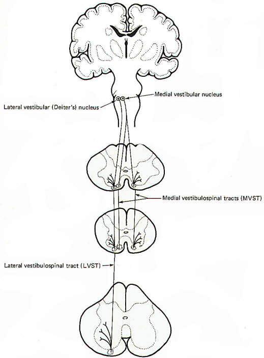
Vestibulospinal Tracts
The
vestibulospinal tracts originate in the vestibular nuclei of the
brainstem. Those fibers originating in the lateral vestibular (Deiter's)
nucleus descend ipsilaterally in the anterior funiculus and form
the lateral vestibulospinal tract (LVST) (Fig-7). The fibers
of this tract terminate in laminae VII, VIII, and IX at all
levels of the cord. The vestibulospinal tracts facilitate
extensor and inhibit flexor alpha and gamma motor neurons.
Input from the vestibular apparatus to the vestibular nuclei
via cranial nerve VIII presupposes an antigravity or postural
role for the lateral vestibulospinal tract. Activity in this
tract is also influenced by input to the vestibular nuclei from
the cerebellum. Arising from the medial vestibular nucleus are
the fibers of the medial vestibulospinal tract (MVST). While
there is a small crossed component, most of its fibers descend
ipsilaterally only as far as the midthoracic cord, where they
too synapse in laminae VII, VIII, and IX. The function of this
tract may be similar to that of the lateral vestibulospinal
tract, but its precise role is largely unknown.
Interstitiospinal Tract
The
descending fibers of this tract arise in the interstitial
nucleus of Cajal (an accessory nucleus of III) in the tegmentum
of the midbrain. They descend ipsilaterally only to the cervical
level of the cord, where they synapse in laminae VI, VII, and VIII. The tract may play a role in reflex movements of the head
and neck in response to visual stimuli, but its function is
largely unknown and probably more complex.
Tectospinal
Tract
The
descending fibers of this tract arise chiefly in the tectum of
the superior colliculus. Some of them decussate and others
don't. In either case they only descend to cervical levels where
they synapse in laminae VI, VII, and VIII. The tract has been
implicated in mediating visual reflexes but, again, its
function is largely unknown.
|
 |
| Fig-7 |
|
|
|
|
|
Prof. Munir Elias
Our brain is a mystery and to understand it, you
need to be a neurosurgeon, neuroanatomist and neurophysiologist.
neurosurgery.tv

Please visit this site, where daily neurosurgical activities are going
on.
Inomed ISIS IOM System
|
|
|
|