|
|
 |
CEREBRAL BLOOD FLOW AND OXYGEN CONSUMPTION
 |
CEREBRAL BLOOD FLOW
The average
cerebral blood flow in humans is approximately 55 mL per
100 g of brain tissue per minute. This is a little over
700 mL/min for a 1350-g brain. Thus while the human
brain comprises only about 2.5 percent of the body's
weight, it receives almost 15 percent of the cardiac
output, attesting to the high vascular demands of this
organ.
A reliable and
frequently used method of determining cerebral blood
flow is the method of Kety and Schmidt. It is based on
the Fick principle and utilizes the arteriovenous
difference of a freely diffusible gas such as N2O
as it passes through the brain. Accordingly, the flow of
blood through the brain can be determined by measuring
the amount of N2O removed from the blood by
the brain per minute and dividing this by the
arteriovenous difference of N2O as it passes
through the brain. The cerebral blood flow is higher in
children than in adults, typically exceeding 100 mL per
100 g per minute. However, contrary to popular thinking,
the blood flow decreases only slightly with advancing
age. The brain utilizes fully 25 percent of the body's
total oxygen consumption. The arteriovenous O2
difference is relatively high since the brain receives
only 15 percent of the cardiac output. The arteriovenous
difference is 6.6 mL per 100 ml., falling from 19.6 to
13 mL per 100 mL as blood passes through the brain (Fig.
17-2). Thus we can calculate a cerebral oxygen
consumption of approximately 3.5 mL per 100 g per
minute. This value is greater in skeletal muscle, skin.
and liver. but less in cardiac muscle and kidney.
|
 |
OXYGEN
CONSUMPTION
The utilization of
oxygen by the brain is not uniform throughout its mass.
The gray matter consumes as much as 94 percent of
cerebral oxygen. while the white matter, which makes up
fully 60 percent of the brain's mass. consumes only 6
percent. Oxygen consumption, and hence oxygen need,
increases as we move up the neuraxis. It is lowest in
the spinal cord and increases through the medulla.
midbrain, thalamus, cerebellum, and cerebral cortex.
Thus it is not surprising to find that the sensorimotor
functions of the cerebral cortex are more sensitive to
hypoxic damage than are the vegetative functions of the
pontomedullary areas. Progressive decreases in cerebral
oxygen consumption are always accompanied by progressive
decreases in the level of mental alertness. Compared to
the mentally alert young man with an O2
consumption of 3.5 mL per 100 g per minute, the mentally
confused states associated with diabetic acidosis,
insulin hypoglycemia, and some forms of cerebral
arteriosclerosis might typically show O2
consumptions rates down to 2.8 mL per 100 g per minute.
Finally, the comatose states of diabetic coma. insulin
coma. and anesthesia can show consumption rates as low
as 2.0 mL per 100 g per minute. On the other hand. O2
consumption by the brain increases during convulsions. |
 |
EFFECTS OF OXYGEN DEPRIVATION
Almost all of the oxygen consumed by the brain is
utilized for the oxidation of carbohydrate.
Sufficient energy is released from this process so
that the normal level of oxygen utilization is
adequate to replace the 12 mmol or so of A TP which
the whole brain uses per minute. However, since the
normal brain reserve of A TP and creatine phosphate
(CrP) totals only about 8 rnmol, less than a
minute's reserve of high energy phosphate bonds is
actually available if production were to suddenly
stop. In the absence of oxygen, the anerobic
glycolysis of glucose and glycogen could supply only
another 15 mmol of A TP, as these two energy
substrates are stored in such low quantities in
brain tissue.
A continuous uninterrupted supply of oxygen to the
brain is essential in order to maintain its
metabolic functions and to prevent tissue damage.
The oxygen-independent glycolytic pathway (anerobic
glycolysis) is insufficient, even at maximum
operating levels, to supply the heavy demands of the
brain. Thus a loss of consciousness occurs when
brain tissue P02 levels fall to 15 to
20
mmHg. This level is reached in less than
10
s when cerebral blood flow is completely stopped
Low tissue oxygen levels in the brain (hypoxidosis)
can be caused by decreased blood flow (ischemia) or
with adequate blood flow accompanied by low levels
of blood oxygen (hypoxemia). It is important to
recognize that decreased P02 caused by
ischemia is accompanied by decreased brain glucose
and increased brain CO2 while hypoxemia
with normal blood flow is not accompanied by changes
in brain glucose or CO2,
with
complete cessation of CBF, irreversible damage
occurs to brain tissue within a few minutes and the
histological effects observed are remarkably
similar whether caused by ischemia, hypoxemia, or
hypoglycemia.
Experimental studies on rats and mice in which
arterial P02 is progressively reduced
have illustrated some aspects of hypoxemia which are
likely to be similar in humans. A drop in arterial
P02
to 50 mmHg (normal, 96 mmHg) produces no change in
CBF, O2
utilization by the brain, or lactic acid
production. However, as P02
levels drop to 30 mmHg, a 50 percent increase in CBF
is observed along with the onset of coma, decreased
oxygen utilization, and increased lactic acid
production. When the P02 drops further to
15 mmHg, 50 percent of the animals die because of
cardiac failure. The remainder show a tremendous
increase in lactic acid production, but,
surprisingly, levels of ATP, ADP, and AMP remain
normal. If cerebral perfusion is artificially
maintained while the arterial P02
is decreased further, ATP, ADP, and AMP levels still
remain normal. The implication is that the coma
observed at low oxygen levels may not be due to a
decrease in ATP but instead to some still
unexplained mechanism. It appears likely that
cardiac complications caused by hypoxemia and the
subsequent effect on cerebral blood flow may
actually be a primary cause of the irreversible
pathologic damage to the brain.
Hypoxia, such as that brought on by high altitudes,
brings on a number of symptoms, including
drowsiness, apathy, and decreases in judgment.
Unless oxygen is administered within half a minute
or so, coma, convulsions, and depression of the EEG
occur.
|
|
 |
 |
|

Prof. Munir Elias
Our brain is a mystery and to understand it, you
need to be a neurosurgeon, neuroanatomist and neurophysiologist.

neurosurgery.tv

Please visit this site, where daily neurosurgical activities are going
on.

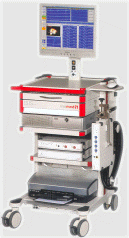
Inomed ISIS IOM System
|
|
|
|